Why You Need Dental X-Rays
Learn how often you should get radiographs and how safe they are
X-rays, also known as radiographs, capture images of the parts of your mouth your dentist can’t see. That’s because hard tissues like bones and teeth absorb more radiation than softer gum and cheek tissues, creating a picture that clearly shows differences between these types of tissues.
Dentist can use X-ray technology to diagnose cavities, gum disease, infection, tooth cracks, bone loss and other problems that aren’t visible to the eye. In addition, X-rays help the dentist find and treat dental problems early in their development, which can potentially save you money and unnecessary discomfort. X-rays play a big part in keeping your teeth and gums healthy.
What Problems Can Dental X-Rays Detect?
Dental X-rays can be used to:
- Show areas of decay that may not be visible with an oral exam, especially cavities between teeth
- Identify cavities occurring under an existing filling
- Reveal bone loss and periodontal disease
- Reveal changes in the bone or in the root canal resulting from infection
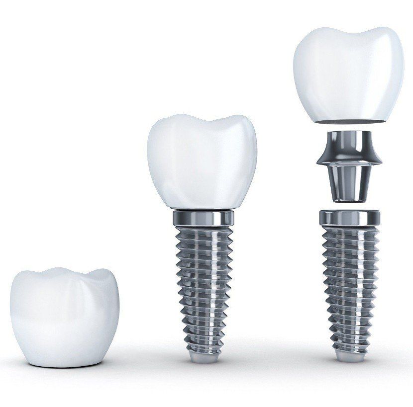
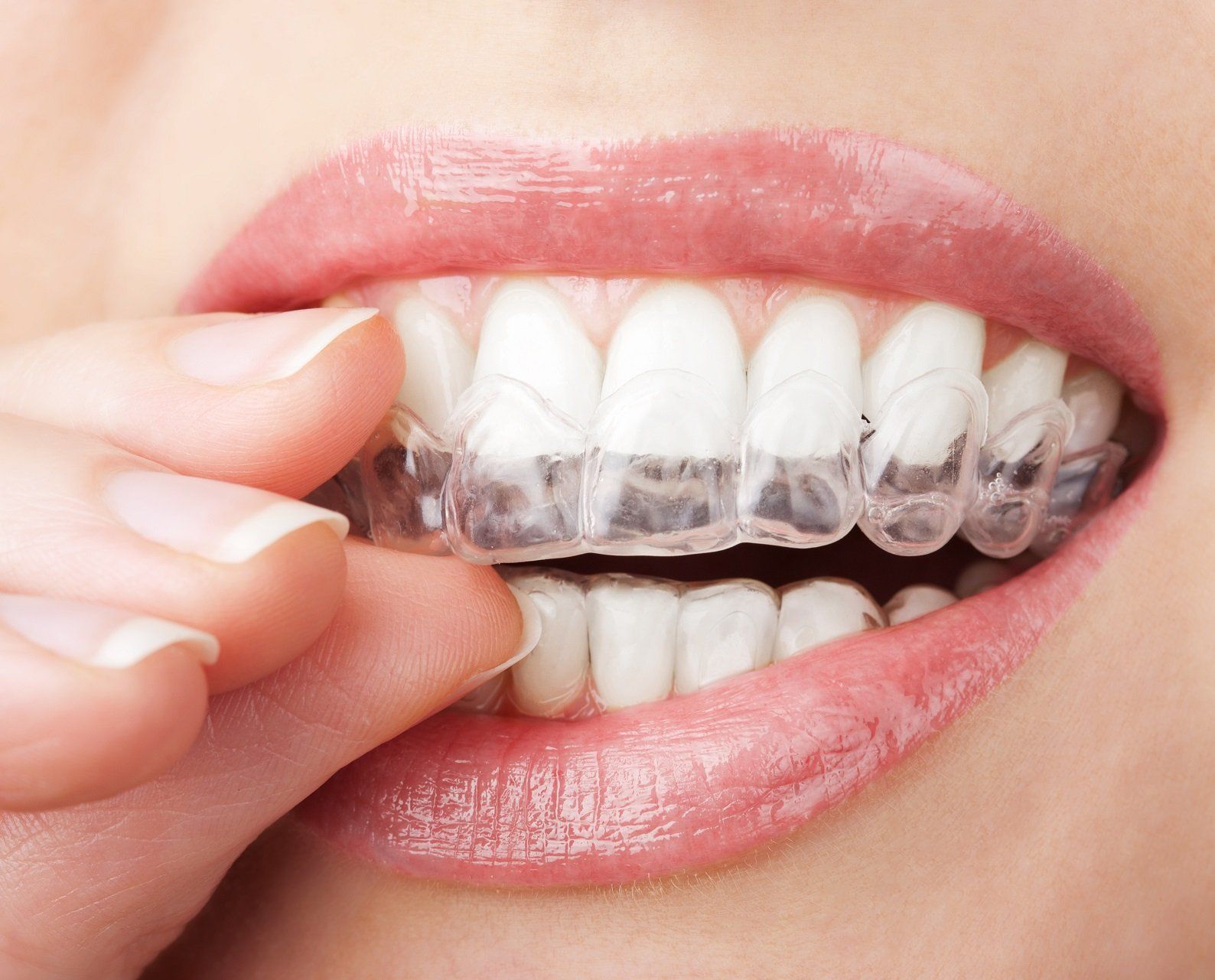
- Assist in the preparation of tooth implants, braces, dentures, or other dental procedures
- Reveal an abscess (an infection at the root of a tooth)
- Reveal other developmental abnormalities, such as cysts and some types of tumors
- Incipient (early stage) decay
- See the status of developing teeth
- Determine if primary teeth are being lost quickly enough to allow permanent teeth to come in properly
- Check for the development of wisdom teeth and identify if the teeth are impacted (unable to emerge through the gums)
How Often Should Teeth Be X-Rayed?
The frequency of getting X-rays of your teeth often depends on your medical and dental history and current condition. Some people may need X-rays as often as every six months; others with no recent dental or gum disease and who visit their dentist regularly may get X-rays only once a year. If you are a new patient, your dentist may take X-rays as part of the initial exam and to establish a baseline record from which to compare changes that may occur over time.
People who fall into the high risk category who may need X-rays taken more frequently include:
- Children generally need more X-rays than adults because their teeth and jaws are still developing and because their teeth are smaller and enamel of their teeth is finer. As a result, decay can reach the inner part of the tooth, dentin, quicker and spread faster.
- Adults with extensive restorative work, such as fillings, crowns and bridges to look for decay beneath existing fillings.
- Adults that wear removable appliance like dentures and partials.
- People who drink a lot of sugary and acidic beverages .
- People with periodontal disease to monitor bone loss.
- People who have dry mouth - called xerostomia - whether due to medications or disease states (such as Sjogren's syndrome, damaged salivary glands, radiation treatment to head and neck). Dry mouth conditions can lead to the development of cavities.
- Smokers to monitor bone loss resulting from periodontal disease.
How Safe Are Dental X-Rays?
Exposure to all sources of radiation -- including the sun, minerals in the soil, appliances in your home, and dental X-rays -- can damage the body's tissues and cells and can lead to the development of cancer in some instances. Fortunately, the dose of radiation you are exposed to during the taking of dental X-rays is extremely small, especially if your dentist is using digital X-rays.
Advances in dentistry over the years have lead to a number of measures that will minimize the risks associated with X-rays. However, even with the advancements in safety, the effects of radiation are added together over a lifetime. So every little bit of radiation you receive from all sources counts.
If you are concerned about radiation exposure due to X-rays, talk to your dentist about how often X-rays are needed and why they are being taken.
Types of Dental X-Rays:
·Bite-Wing x-rays show both lower and upper back teeth in one x-ray. These types of x-rays are used to diagnose in-between and chewing surface cavities. They are also used to evaluate bone around the teeth and aid in diagnosing periodontal disease.
·Panoramic x-ray shows a broad view of the teeth, entire jaws, nasal area, sinus and large lower jaw nerves. This type of x-rays are usually used for extractions, implant placements, locating and evaluating 3rd (wisdom) molars, looking for any abnormalities (cysts or abscesses) and when going thru orthodontic treatments.
·Periapical x-rays provide a view of the entire tooth, from the crown to the bone that helps to support the tooth. These type of x-rays are used to find dental problems below the gum line or in the jaw, such abscesses, cysts, and bone changes.
·3D images or Cone Beam Imaging is a diagnostic imaging technology that uses radiation in a manner similar to conventional radiographic imaging, with the difference being that cone beam images are converted into a three-dimensional view that can then be manipulated by computer software for a wide variety of applications, including implant, orthodontic, TMJ, and diagnostic purposes.
For more information, please contact us at your convenience by calling 330-733-7911 or sending us a website message.
